First Degree AV Block
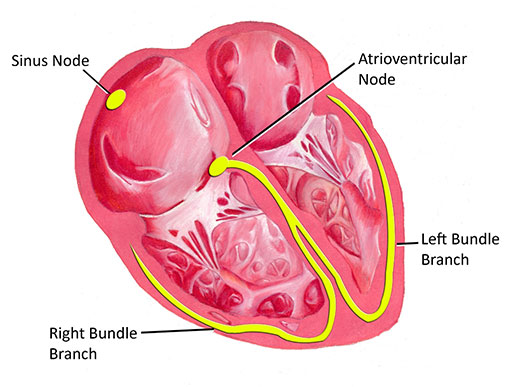
The AV node is a group of specialized conducting cells found in the middle of the heart. The AV node is responsible for transmitting electrical pulses from the top part of the heart to the bottom part of the heart. First-degree AV block refers to a condition where the transmission of impulses through the AV node is slower than expected.
First degree AV block is usually diagnosed incidentally on an electrocardiogram. The typical finding is a prolonged PR interval. The PR interval simply measures the time it takes for electricity to move through the atrium into the ventricles. A longer than normal transit time is usually due to slow conduction through the AV node.
In many instances first degree AV block is a completely normal finding caused by increased activity of certain nerves of the body. For example, the vagus nerve, a large nerve that travels from the brainstem throughout the body has branches that go to the AV node. When the vagus nerve is activated, transmission of electricity through the AV node may be slightly slower than normal, producing first-degree AV block. Many completely healthy individuals may show evidence of first degree AV block during times of relaxation or sleep.
First-degree AV block may also be a sign of potential AV node dysfunction. Some patients may have progressive electrical conduction system disease that initially manifests as first degree AV block.


First-degree AV block in and of itself does not produce any symptoms. Evaluation usually includes an electrocardiogram (ECG); in some instances a more prolonged recording of the heart rhythm is necessary (24-hour Holter monitor). Finally, in certain cases a stress test may be helpful to evaluate electrical conduction during exercise.
First-degree AV block generally does not require any treatment. If it is thought to be physiologic or a normal variant, further follow-up may not even be necessary. If there is any concern for more diffuse electrical conduction disease, then more frequent reevaluation is usually required.
