- Details
Your baby came to the cardiologist for evaluation of a heart murmur and the doctor tells you that your newborn has mild PPS, peripheral pulmonary stenosis. Then they tell you that this is a common finding in newborn babies and that you shouldn't worry. But it sounds terrifying! So what is it?
Peripheral pulmonary stenosis, PPS, is the term used to describe narrowing of the arteries that take de-oxygenated (blue) blood from the right ventricle to the lungs. Some doctors may also call it branch pulmonary artery stenosis, because it occurs at the branch points of the main pulmonary artery.
- Details
Atrial fibrillation is the most common arrhythmia found in older adults. Most people have heard of it before. In fact, most people either have a relative, parent, or other friend that has experienced atrial fibrillation. It's just that common!
- Details
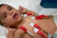
Influenza (“the flu”) is a contagious respiratory illness cause by influenza viruses. When a person contracts the flu virus, their Illness can vary from mild to severe and can lead to hospitalization and sometimes even death. There are some specific types of people that are at a higher risk for developing serious complications from the flu. Those include the elderly, young children and people with certain medical conditions.
- Fever, cough, sore throat, runny or stuffy nose, body aches, headache, chills and fatigue
- Some people also experience vomiting and diarrhea
- Some people may have all the upper respiratory symptoms without a fever
The flu vaccine comes in various forms - either by a shot or a nasal spray. The vaccine causes antibodies to develop in the body about two weeks after it is administered. Every year the vaccine protects against the three viruses that research indicates will be the most common during the upcoming season. This year the vaccine includes influenza A (H1N1) virus, influenza A (H3N2) virus, and a type of influenza B virus. The flu vaccine should be administered every year because the vaccine is formulated on a yearly basis as the flu virus changes over time.
The Flu Shot:
an inactivated vaccine containing a killed form of the virus that is given with a needle in the arm. The flu shot is approved for any person six months of age and older. There are three forms of the shot:
- Regular flu shot: approved for people ages six months and older
- High dose flu shot: approved for people 65 years and older
- Intradermal flu shot: approved for people ages 18 to 64
-
Side effects from the flu shot:
- Soreness, redness or swelling where the shot was given.
- Low-grade fever
- Muscle aches
The Nasal-Spray Flu Vaccine
made with live, weakened flu viruses (live-attenuated influenza vaccine “LAIV”) given in the form of a spray in the nose. This form of the vaccine does not cause the flu. This form of the vaccine is approved for healthy people ages 2 to 49 years who are not pregnant.
Side effects of the nasal-spray:
- Runny nose
- Wheezing
- Headache
- Vomiting
- Muscle aches
- Difficulty breathing, hoarseness or wheezing.
- Swelling around the eyes or lips
- Hives
- Paleness
- Weakness
- Fast heart beat
- Dizziness
- High fever
- Behavioral changes
Every person over the age of 6 months should receive the flu vaccine. It is especially important for people who are at a high risk of developing complications from the flu to receive the vaccine. People who live and care for individuals in this high risk population should be vaccinated as well.
High-risk populations include:
- People with certain medical conditions including: Heart disease, asthma, diabetes, chronic lung disease, liver disease, blood disorders
- Pregnant women
- People 65 years of age and older
- People aged 6 months to 18 years and are on long-term aspirin therapy
All of our physicians are experts in the field of congenital heart disease diagnosis, evaluation, and management. Some of the more complex forms of heart disease that we treat include hypoplastic left heart syndrome, tetralogy of Fallot, transposition of the great arteries, pulmonary valve atresia, pulmonary valve stenosis, aortic valve stenosis, atrioventricular canal defect, ventricular septal defect, atrial septal defect, Ebstein’s anomaly, and coarctation of the aorta, as well as many other forms of heart disease. We are also experts in heart rhythm disorders such as supraventricular tachycardia (SVT).
We will provide informative and compassionate counseling to you and your family. When necessary, we will arrange for further consultation with a congenital heart surgeon should you desire.
All of our physicians are experts in fetal cardiac diagnosis and management. We work closely with your obstetrician and perinatologist to assist in the management of your child. We have extensive experience with all forms of complex congenital heart disease, including hypoplastic left heart syndrome, tetralogy of Fallot, pulmonary valve atresia, aortic stenosis, transposition of the great arteries, atrioventricular canal defect, ventricular septal defect, Ebstein’s anomaly of the tricuspid valve, and coarctation of the aorta, as well as many other forms of heart disease. We are also experts in heart rhythm disorders such as supraventricular tachycardia (SVT) and congenital complete atrioventricular block.
We provide informative and compassionate counseling to you and your family. When necessary, we can arrange for further consultation with a congenital heart surgeon.
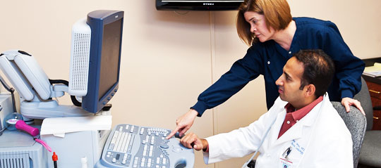
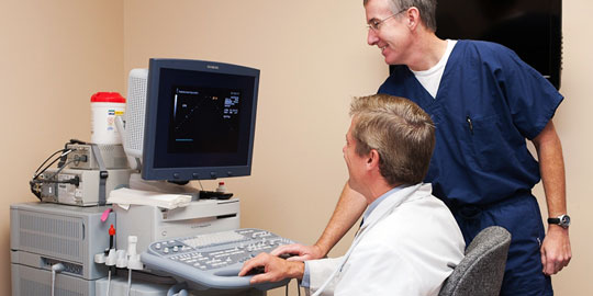
A fetal echocardiogram is a detailed study of the heart of a developing fetus. Your doctor may request a fetal echocardiogram for a number of different reasons. These may include suspicion of a congenital heart defect, a family history of congenital heart disease, or certain conditions in the mother which may predispose to heart disease such as diabetes or phenylketonuria. A fetal echocardiogram can usually be performed successfully after 18 weeks gestation, occasionally sooner.
The physicians at Pediatric Heart Specialists are experts in fetal echocardiogram perfomance. All our locations throughout Dallas and East Texas have the capability to perform fetal echocardiograms.
Your fetal echocardiogram will be performed in a private room by one of our physicians. Once the study is complete, the doctor will spend as much time as is necessary reviewing the results with you and providing in-depth counseling. We understand that this can be an extremely stressful time for you. Our physicians and office staff will treat you with compassion and understanding and will work hard to make your visit as informative as possible.
- Details
As a parent, having your child complain of chest pain can be a scary concept.
It is natural to worry about the heart first. Fortunately, while chest pain
in adults can often be cardiac, most chest pain in children is not related to
the heart.
There are a number of non-cardiac factors that can lead to chest pain in
children. The most common cause of chest pain in children and teenagers is
chest wall pain. Typically a brief, sharp sensation, this pain is sometimes
worsened with deep breathing. This is a benign cause of pain, generally
thought to be secondary to muscle spasms within the chest or irritation of
the lung pleura (lining). While this pain is not caused by the heart,
it is real pain and can cause distress to the child.
- Details
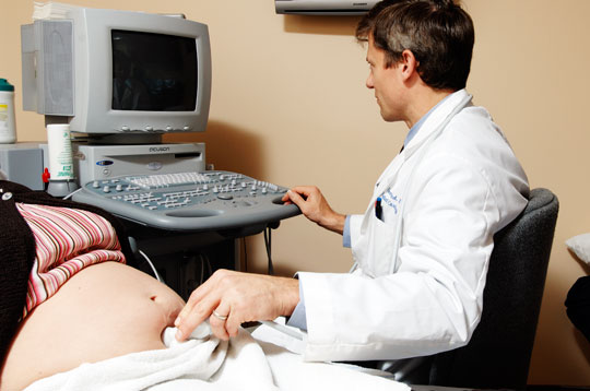
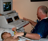
A fetal echocardiogram is a detailed ultrasound of the baby’s heart performed before the baby is born. It is a more comprehensive evaluation of the heart than what is done during the typical obstetrical ultrasound. A fetal echocardiogram can evaluate both the structure and function of the heart. For this reason it is useful in looking for birth defects of the heart and heart rhythm problems as well as for assessing fetal well being if the baby is found to have other potential problems during the pregnancy.
There are three categories of risk factors that should prompt fetal cardiac evaluation with a fetal echocardiogram: maternal risk factors, fetal risk factors, and family risk factors. Maternal risk factors include a mother who has taken medications that are known to cause congenital heart defects, a mother who has specific health problems (such as diabetes or an autoimmune disease), or a mother who has had certain infections during the pregnancy (such as rubella or CMV). Fetal risk factors include a fetus that has been diagnosed with a genetic abnormality (such as Down syndrome) or has had an abnormal amniocentesis, a fetus who has abnormalities in other organs (such as the brain, kidney, or GI tract), a fetus with an abnormal heart rate or rhythm (too fast or too slow), or a fetus in whom a heart abnormality is suspected on a routine ultrasound. Familial risk factors include a first degree relative (father, mother, or sibling) with a congenital heart defect or a known family history of disorders that are passed from generation to generation (such as Marfan syndrome, DiGeorge syndrome, or tuberous sclerosis).
A fetal echocardiogram is generally performed by a songographer in conjunction with a pediatric cardiologist specialized in fetal congenital heart disease. A fetal echocardiogram can be performed any time after 17-18 weeks gestation, though the images are usually optimal in the 20-24 week range (a transvaginal fetal echocardiogram can be performed as early as 12-13 weeks, but this approach is not commonly used). A typical exam takes from 30-90 minutes.
Following the fetal echocardiogram, the cardiologist will sit with you and explain in detail (with pictures and diagrams) the findings of the test and what it means for you and your baby. If a congenital heart defect is identified, we will do our best to make sure you understand every aspect of the diagnosis and how it will affect the baby before delivery, during delivery, and throughout your child’s lifetime.
Dr. Tracy Laird
Posted by .